
Echocardiography (Cardiac Ultrasound)
Echocardiography is used to evaluate many heart functions, such as the size of the heart, the size of the heart chambers, the thickness of the heart muscle, valve function, blood flow velocity, and the condition of the blood vessels. This process is essential for the diagnosis and treatment of heart diseases. It is also used for follow-up before and after heart surgeries. It is a completely painless, fast and safe procedure.
What is Echocardiography?
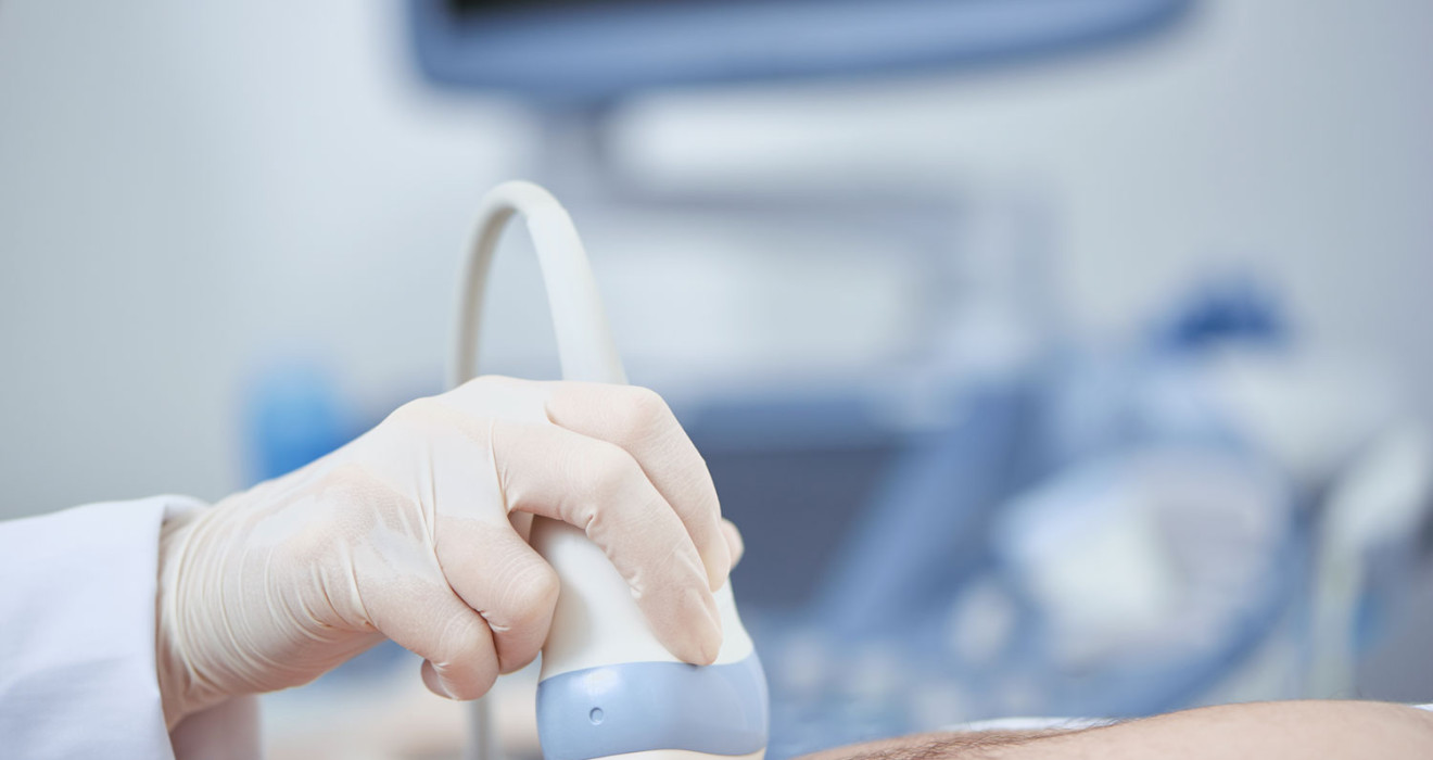
Echocardiography is the process of imaging the heart using ultrasound waves. An ultrasound device and a probe are used in this process.
The probe is placed over the heart, and the ultrasound waves are reflected from the heart. The computer processes these waves to create real-time heart images.
When to Get Echocardiography?
Cardiac ultrasound is used in many different situations. The main conditions in which it is used are as follows:
- Diagnosis of heart diseases
- After a heart attack
- Vascular occlusions
- Before heart surgery
- Genetic heart diseases
- After heart surgery
- Hypertension
What are the Types of Echocardiography?
Types of echocardiography are as follows:
- Transthoracic (TTE): It is the most common type of echocardiography. It is an imaging procedure performed outside the heart using ultrasound waves.
- Transesophageal (TEE): It obtains ultrasound waves near the heart by going down the oesophagus with an endoscopic probe.
- Stress echocardiography: It is used to evaluate heart functions under stress factors such as exercise or drug use.
- 3D heart ultrasound: Based on the same principle as TTE or TEE. However, it uses three-dimensional imaging to obtain a more detailed image.
- Contrast-enhanced cardiac ultrasound: A contrast agent is administered intravenously. This substance is used to show the blood in the heart cavities clearly.
- Speckle tracking cardiac ultrasound: It is used to measure the movement of the heart muscle.
How Is Echocardiography Taken?
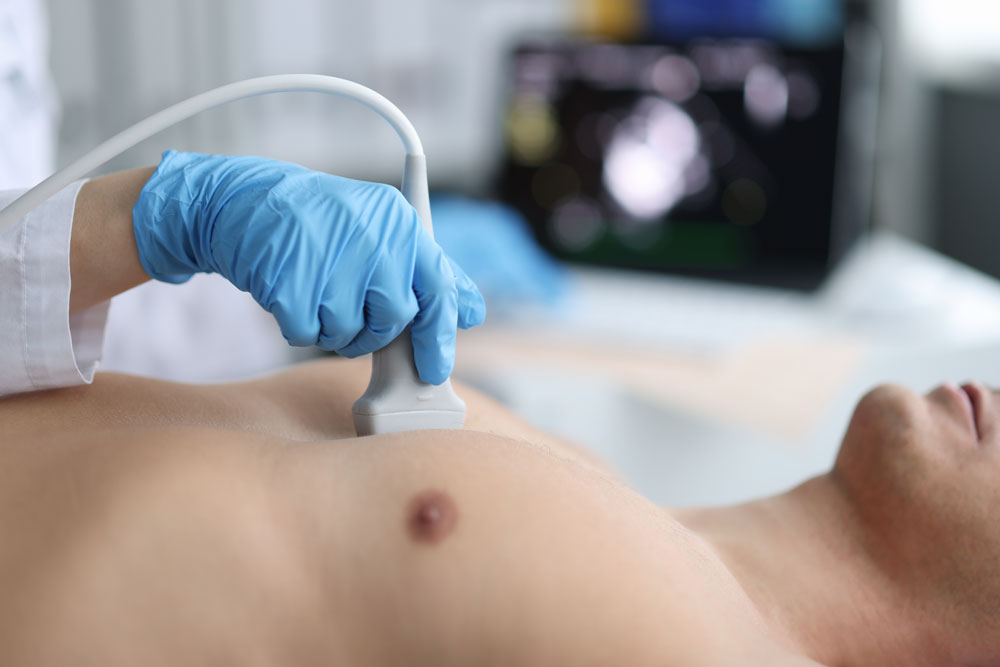
Echocardiography is performed using an ultrasound device and a probe, usually on a table or bed with the patient lying on their back.
During the procedure, a gel is applied to the patient's chest to provide a better image of the probe. The image of the heart is viewed on the monitor, and details such as heart muscle movements, heart valves and blood flow rate are examined.
The cardiac ultrasound procedure is usually painless and quick. It takes about 5-15 minutes. Results are generally immediately available and interpreted.
What Does Echocardiography Show?
The echocardiography shows the following:
- Heart structure
- Blood flow velocity and direction
- State of cardiac muscle functions
- Cardiac valve diseases
- Blood clots
What is the Difference Between ECG and ECHO?
ECG (Electrocardiography) and ECHO (Echocardiography) are medical tests that provide information about heart health. However, they provide different information.
ECG measures the electrical activity of the heart. It is used to determine whether the heartbeat is regular or irregular, whether the left and right sides of the heart work equally, and whether there is damage to the heart muscle in any heart attack.
ECHO measures the structural features of the heart. It is used to evaluate the heart muscle's thickness, the heart chambers' size, the heart valves' function, and the heart's overall pumping function.
Frequently Asked Questions
-
Cardiac ultrasound does not show vascular occlusion.
-
The results are presented in a report showing parameters such as the size and shape of the heart, valve functions, blood flow rate and muscle strength of the heart. The doctor reviews these results.
-
The EF (ejection fraction) value measures how much blood is pumped through your heart with each beat. Normal EF value is usually between 55-70%.
-
Echocardiography usually takes between 5-15 minutes.
-
Cardiac ultrasound can be applied to many patients with heart problems or who need information about heart functions. However, in some cases, it is not possible to take images of the heart using ultrasound waves. These conditions include extreme obesity or bone anomalies in the rib cage.
-
Your doctor decides whether you should get ECHO.