
Coronary Tomographic Angiography (Virtual Angiography)
Tomographic angiography is considered a less risky and less uncomfortable procedure because it is non invasive. In this method, a special dye (contrast medium) is injected into a blood vessel in the arm or wrist area of the patient using a catheter or needle. A computed tomography (CT) scanner then uses special software to image the blood vessels inside the body.
What is Tomographic Angiography (CT Angiography)?
Tomographic angiography (CT angiography) is a medical imaging method used to visualize blood vessels in the body. It uses computed tomography (CT) technology to identify narrowing or blockages, especially in the heart and coronary arteries.
Why is Tomographic Angiography (CT Angiography) Performed?
CT angiography is widely used in the diagnosis of heart and vascular diseases. It is particularly useful in the diagnosis and treatment of coronary artery disease, aortic aneurysm, peripheral artery disease, pulmonary embolism and other vascular diseases.
How is Virtual Angiography (CT Angiography) Performed?
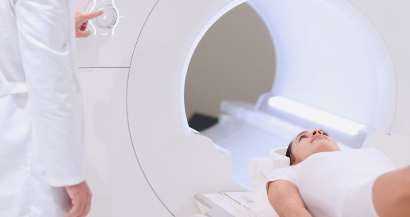
Virtual angiography is a procedure using a series of CT images. First, the patient is injected with a radioactive substance through a venous catheter. This radioactive material travels through the veins during the CT scan, making the images clearer.
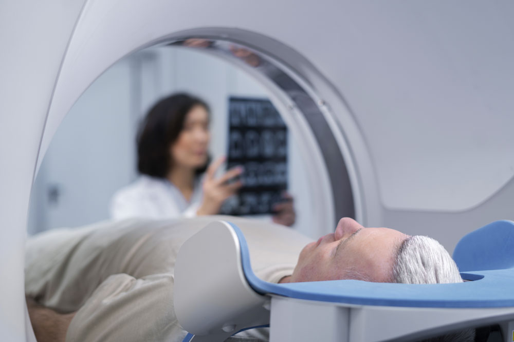
During tomographic angiography, the patient must lie on their back or stomach. The CT scanner scans specific areas of the body in thin slices and the computer converts these slices into a three-dimensional image. These images are analyzed using computer programs and the vessels are visualized.
Who is Tomographic Angiography (CT Angiography) Performed?
Virtual angiography is used to visualize various vessels, such as heart vessels, brain vessels, kidney vessels, lung vessels and leg vessels. It is also performed on patients at risk of coronary artery disease or heart attack.
What are the Differences Between Virtual Angiography and Normal Angiography?
The differences between virtual angiography (tomographic angiography) and normal angiography are as follows:
- Normal angiography is an invasive procedure using a catheter inside an artery. During this procedure, a catheter is advanced through the artery and contrast material is injected. Tomographic angiography, on the other hand, is a non-invasive procedure in which contrast material is injected through a vein in the arm or wrist area without using a needle or catheter.
- Regular angiography can be associated with risks such as bleeding, infection, thrombosis, vascular damage and anesthesia. Virtual angiography has fewer risks because it is non- invasive.
- Normal angiography is a procedure using local anesthesia. Tomographic angiography, on the other hand, is usually painless except for a slight sensation of warmth for a few seconds during the injection of the contrast medium. Therefore, local anesthesia is not required.
- Normal angiography provides more detailed images. Virtual angiography, on the other hand, uses imaging from outside the vessel, so the details vary depending on the thickness of the vessel.
- Normal angiography is used to diagnose heart and vascular diseases. Virtual angiography can be used to visualize all vessels other than the heart.
Frequently Asked Questions
-
People who are not suitable for tomographic angiography are as follows:
- Pregnant women
- Those with severe kidney disease
- Allergic to contrast media
-
Tomographic angiography results are usually available within a few days. However, the exact time when the results will be ready may vary depending on the policies of the institution or hospital performing the test.
-
The duration of the procedure may vary depending on the patient's condition and the size of the treated area. Generally, a full body angiography requires 20 - 30 minutes. However, an angiography performed on a smaller area may take less time.
-
Virtual angiography can provide accurate results. However, just like normal angiography, false positive or false negative results may occur. Therefore, the results should be viewed by an experienced radiologist or cardiologist for accurate interpretation.
-
Tomographic angiography contains contrast material. In some people, it can cause a side effect known as contrast-induced acute kidney injury. Therefore, people at risk of kidney disease, such as those with severe kidney disease, diabetic nephropathy and high blood pressure, should be cautious about the use of contrast media during virtual angiography.
-
Modern medical technologies, such as tomographic angiography, are designed to reduce the use of contrast agents and minimize the risk of damage to the kidneys. In particular, virtual angiography using high-tech contrast agents can reduce the risk of kidney damag